Endothelial Cell Culture Under Shear Stress Using Omi
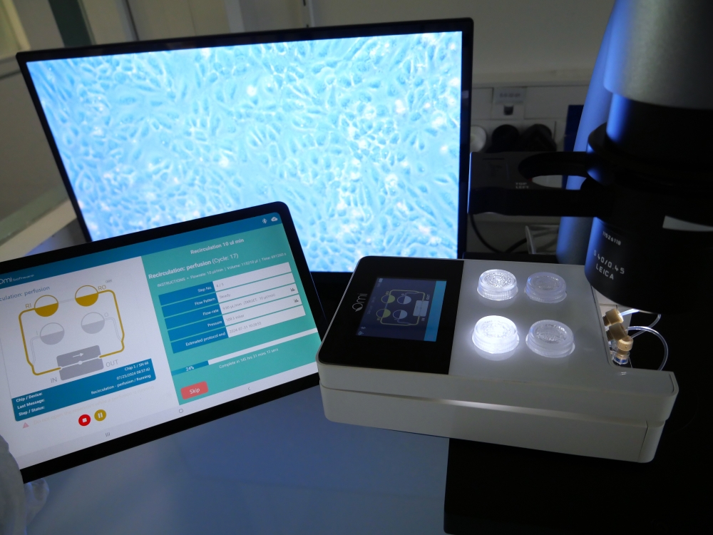
This Application Note provides a protocol for creating a HUVEC monolayer in the BeonChip BeFlow microfluidic chip. After coating and cell seeding, the monolayer is subjected to unidirectional, laminar shear stress using the Omi organ-on-chip automated platform.
Endothelial Cells for Organ-on-Chip Applications
Human umbilical vein endothelial cells (HUVECs) are an extensively used model in organ-on-chip applications. They exhibit key characteristics of vascular endothelial cells, including monolayer formation, extracellular matrix production, and response to angiogenic factors (1). They effectively mimic human vascular endothelium. Their in-vitro use provides a valuable tool for studying vascular endothelium interactions. Organ-on-chip (OoC) technology recreates complex human organ microenvironments in vitro, using microfluidic devices to precisely control and manipulate cellular and biochemical conditions.
Many OoC models can benefit from a controlled endothelial cell culture under shear stress protocol.
- Vascularized Kidney-on-Chip: create a vascular network within kidney-on-chip models.
- Blood-Brain Barrier Models: form part of the endothelial layer in blood-brain barrier models on chips.
- Lung-on-Chip: simulate the vascular compartment of lung-on-chip models.
Shear Stress Effect on Endothelial Cell Culture
Culturing endothelial cells under perfusion in microfluidic systems replicates the dynamic environment of blood vessels, essential for studying endothelial functions. HUVECs are crucial for simulating blood vessel formation in organ-on-chip systems, allowing researchers to investigate blood flow dynamics, shear stress, and endothelial barrier properties (2).
Exposing cells to flow in microchannels helps study the effects of mechanical forces including shear stress on cell morphology, gene expression, and function OoC cell culture under shear stress enhances cell viability, differentiation, and functionality through continuous nutrient supply and waste removal.
Materials & Methods
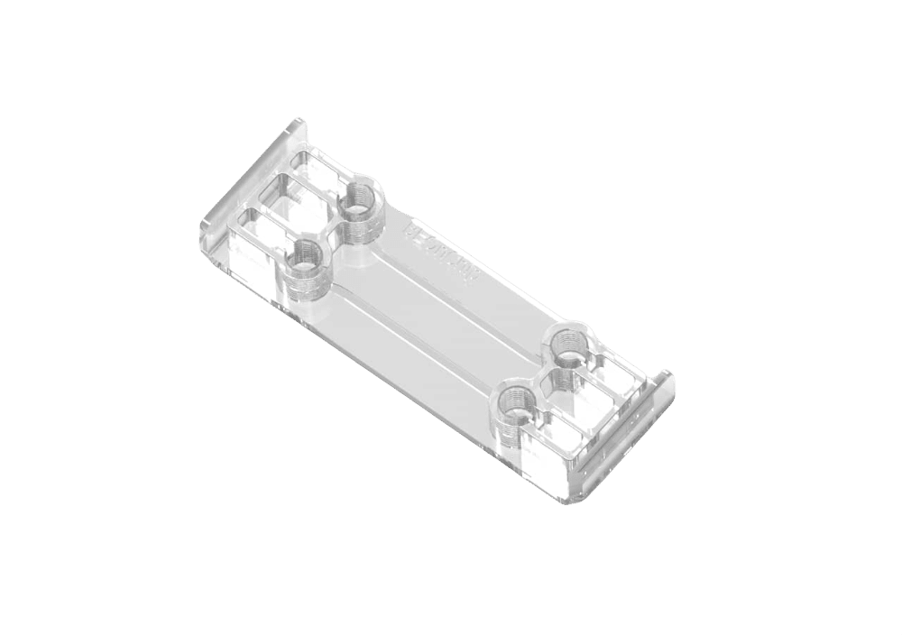
- Microfluidic chip: For this application note, we used the BeonChip Be-flow microfluidic chip (Fig. 1). The Omi is a versatile tool that could be used with any kind of commercially available chip, as well as homemade chips. The channels of the chip are coated with fibronectin protein to enhance cell adhesion (check PDF file for detailed protocol).
- Human umbilical vein endothelial cell culture and seeding: Human Umbilical Vein Endothelial Cells (HUVECs) were sourced from (Lonza, Basel, Switzerland,). They were cultured in Endothelial Cell Growth Medium-2 at 37 ◦C and 5% CO2 up to 7 passages. Detailed protocol is to be found in the PDF.
- Omi setup: The Omi is an advanced automated OOC platform designed for precise liquid flow control in organ-on-chip applications. It is highly accurate and versatile and is compatible with a wide range of microfluidic chips, tubing and connectors.
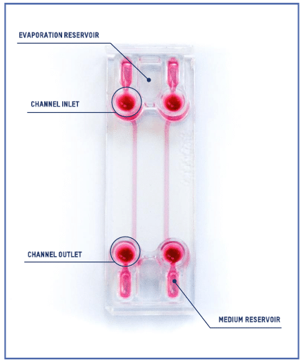
Figure 1: Be-Flow microfluidic chip (BeOnChip)
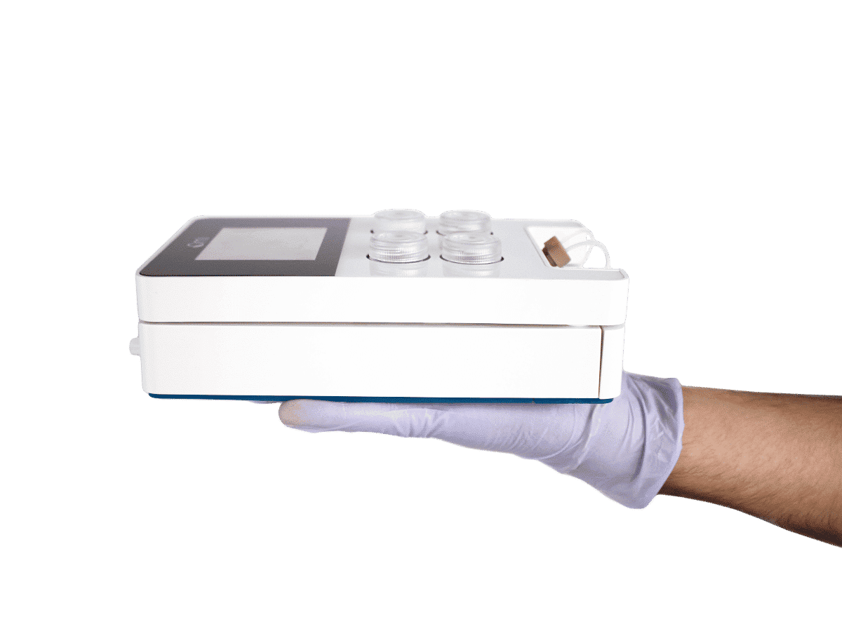
Figure 2: The Omi, automated OoC platform.
Endothelial cell culture under shear stress protocol:
- Sterile conditions: perform all steps under a biological cabinet.
- Attach a sterile reservoir and a low resistance adaptor to the Omi.
- Connect the fluidic circuit and calibrate sensors with sterile water.
- Run the “Sterilization” protocol.
- Replace the low resistance adaptor with a high resistance adaptor.
- Run the “Loading” protocol with the corresponding culture medium to prime the tubings.
- Fill the reservoir with 3 mL of fresh medium.
- Select the desired perfusion channel and ensure the cell monolayer is healthy.
- Wash cells gently with fresh culture media.
- Add fresh medium to the inlet, tilting the chip for slow flow, and fill the inlet/outlet until a convex meniscus forms.
- Load the “recirculation” protocol for 10 µL/min flow for seven days.
- Incubate the Omi at 37°C.
- After 4-5 hours, pause and change the medium to remove non-adherent cells.
- Replenish culture medium daily, pausing the flow each time.
- Regularly check the cell monolayer under a microscope for morphology, alignment, and confluence.
- Use immunostaining to assess cell integrity and junction formation if needed.
Results
Endothelial cell culture under shear stress was sustained for at least seven days.
As observed in the images (Fig.3), the cells exhibit healthy morphology, continue to proliferate and are oriented in the direction of the flow.
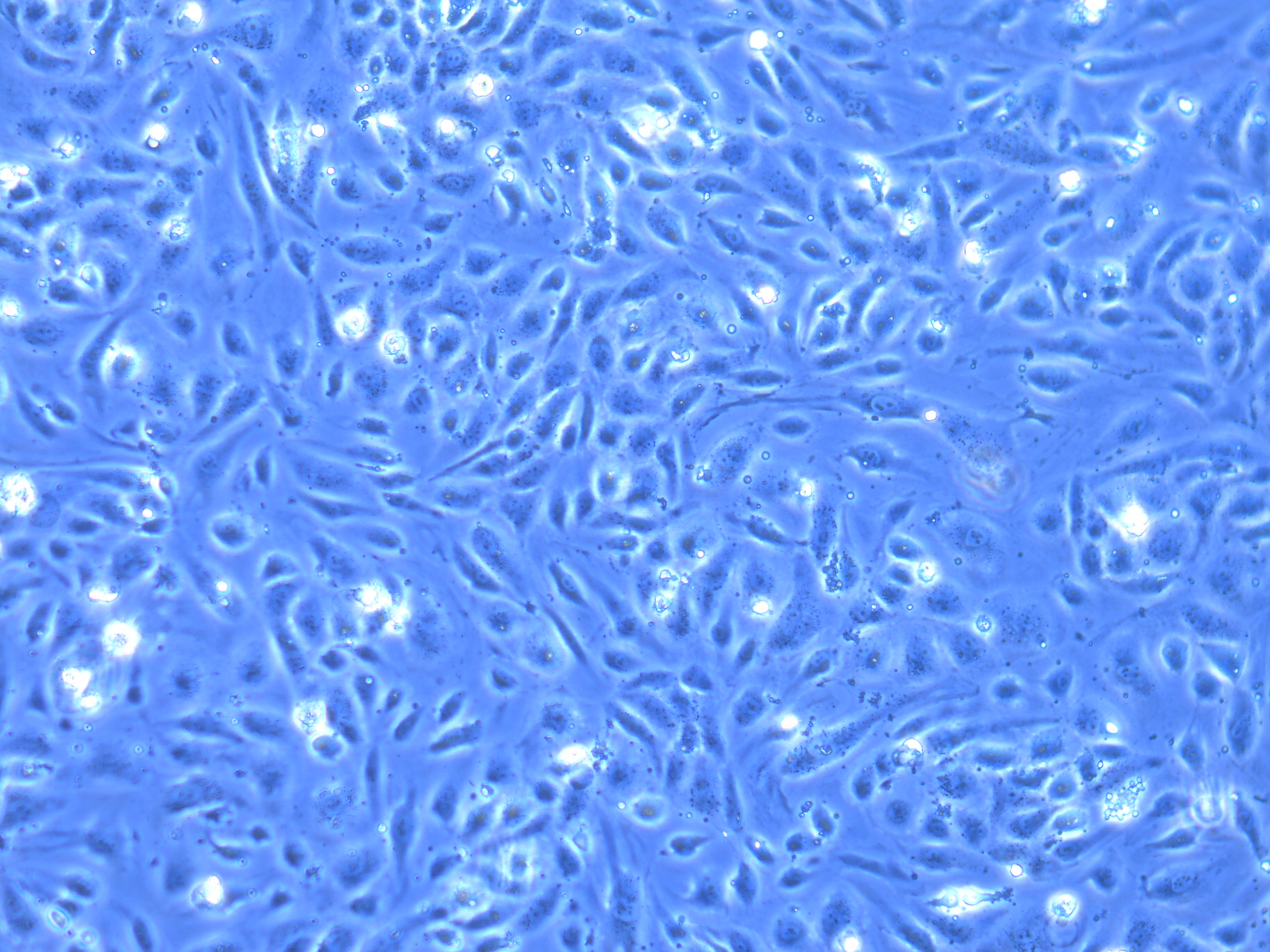
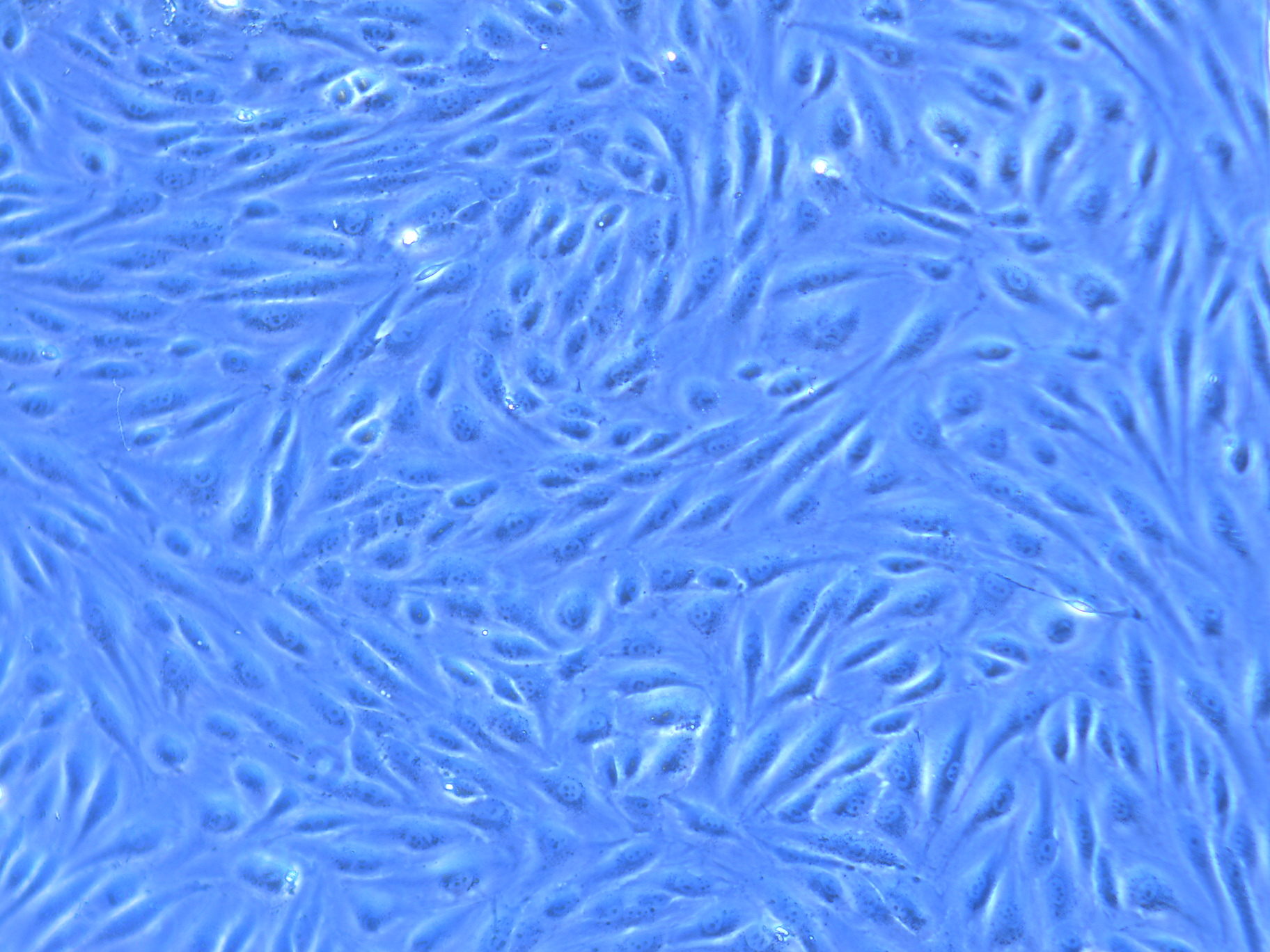
Figure 3: Endothelial cell culture under perfusion, at day 0 and day 7 after perfusion.
Conclusion
Endothelial cell culture under shear stress in microfluidic devices offers a physiologically relevant platform for studying cell biology, disease mechanisms, and therapeutic interventions.
The Omi, our automated (OOC) platform, provides an ideal solution for long-term recirculation of cell culture media in various cell models. It ensures high-precision flow, facilitating the development of a robust monolayer suitable for organ-on-a-chip applications. Easy-to-use, autonomous, and portable under a microscope, Omi is the perfect companion for on-chip biology experiments.
Omi
Omi is an automated platform that helps reproduce the microphysiological behavior of organs inside microfluidic chips. It is compatible any type of chips to sustain different cell culture types or organ on chip models (Liver, Gut, Skin…).
Expertises & Resources
-
Microfluidic Application Notes Omi, automated organ-on-chip platform, the Gut-on-chip model development Read more
-
Microfluidics case studies Creating kidney organoids‑vasculature interaction model using Fluigent’s Flow-EZ Read more
-
Microfluidics case studies A multiplex microfluidic circuit for blood vessel-on-a-chip perfusion using Fluigent’s FlowEZ Read more
-
Expert Reviews: Basics of Microfluidics Microfluidic pressure control for organ-on-a-chip applications: A comprehensive guide Read more
-
Microfluidic Application Notes Long-term fluid recirculation system for Organ-on-a-Chip applications Read more
-
Microfluidics Article Reviews Human Blood Brain Barrier (BBB) permeability -on-chip assessment Read more
-
Expert Reviews: Basics of Microfluidics Why Control Shear Stress in Cell Biology? Read more
-
Microfluidics White Papers An exploration of Microfluidic technology and fluid handling Read more
References
- The use of human umbilical vein endothelial cells (HUVECs) as an in vitro model to assess the toxicity of nanoparticles to endothelium: a review, Yi Cao et al, 2017
- Perfusion culture of endothelial cells under shear stress on microporous membrane in a pressure-driven microphysiological system, Shinji Sugiura et al, 2023.
