Simultaneous labeling of up to 40 proteins within a single tissue section is possible with the IBEX method, also known as “iterative bleaching extends multiplexity”.
This webinar covers this exciting method providing insights into complex biological systems and disease mechanisms.
Meet Dr. Colin Chu from the UCL Institute of Ophthalmology, UK. He played a pivotal role in developing the IBEX technique at the National Institutes of Health, USA.
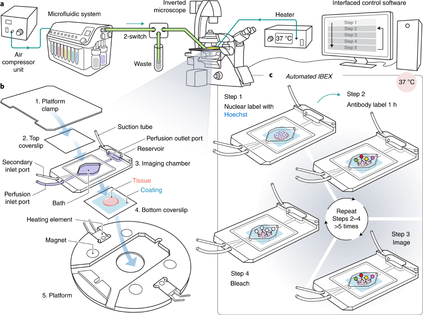
During this webinar, he will outline how they integrated the Fluigent ARIA microfluidic system with a Leica Thunder microscope to automate the process. He will provide an overview of IBEX, its setup, his experience of the system and how research groups can establish this in their own labs.
Date: Wednesday 6/26/2024
Another great news! An IBEX Imaging Community has been created recently! Make sure to join a vibrant network of researchers and professionals dedicated to imaging science and discover their comprehensive knowledge base, exchange insights, and collaborate on pioneering projects!
Join IBEX imaging community here: IBEX Imaging Community
Chu Lab website: https://colinchulab.com/
UCL Chu Lab website: https://iris.ucl.ac.uk/iris/browse/profile?upi=CHUXX55
General UCL website: https://www.ucl.ac.uk/
Automated Perfusion System for Spatial Omics
As a researcher performing live-cell imaging & labeling experiments, you know how they can be expensive (reagents, sample, microscope), time-consuming and stressful. A mistaken operation can ruin the whole experiment.
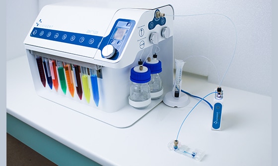
Helping researchers to avoid those risks, to gain productivity, and to get the best of each experiment, Fluigent has created Aria.
Fluigent’s Aria System automates numerous protocols that repetitively stain and image various cells, tissues, and nucleic acids including FISH, DNA Paint, and immunofluorescent protocols.
Adaptable to virtually any microscope, the system allows users to program a sequence of up to ten unique reagents to be delivered to a closed chamber for automated imaging.
IBEX: an iterative immunolabeling and chemical bleaching method for high-content imaging of diverse tissues
Understanding cell-cell interactions and spatial relationships in healthy or diseased organs represents a technical challenge. Single-cell genomics is powerful but lacks spatial context. In this study, automated immunolabeling has been applied to obtain a versatile multiplex optical imaging approach for deep phenotyping and spatial analysis of cells in complex tissues.

